
Ultrasound-guided vascular access is often taught with a strong focus on finding the vessel and maintaining needle visualization. But successful vessel identification is only part of the assessment.
What surrounds the vessel matters too.
In Don’t Get on My Nerves, Wayne Teipen highlights an important anatomical consideration when accessing the basilic vein: the medial antebrachial cutaneous nerve (MACN). The MACN travels adjacent to the basilic vein, and one or more branches may lie directly within a potential needle trajectory.
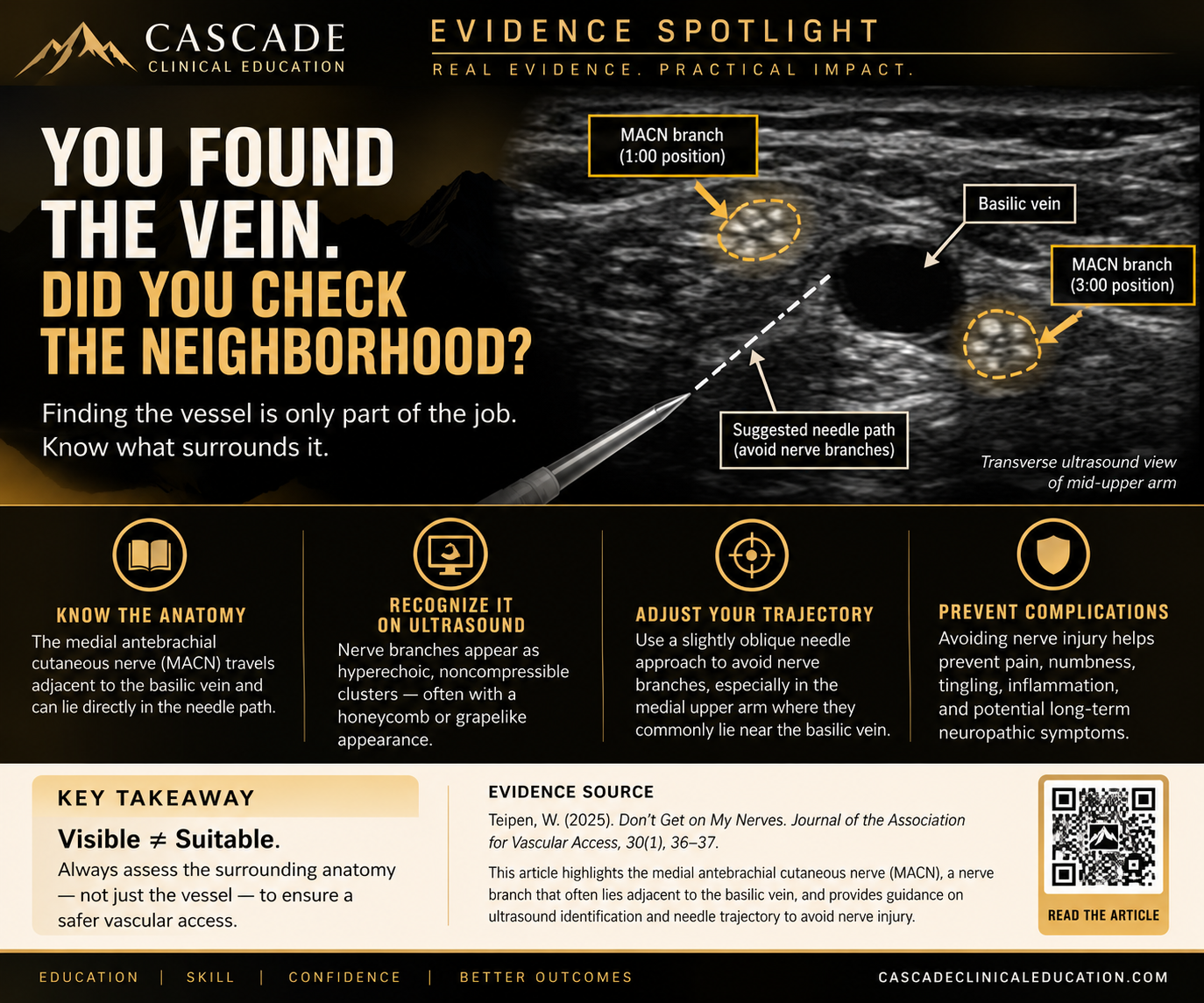
Structure recognition is necessary
Why this matters
Historically, inadvertent nerve contact during vascular access has often been recognized only after a patient reports a sudden electrical or “shocking” sensation. That feedback isn't always reliable—or even possible—in patients who are sedated, anesthetized, unable to communicate effectively, or very young. Ultrasound provides an opportunity to move from reacting to nerve contact to identifying potentially vulnerable anatomy before puncture.
What does a nerve look like on ultrasound?
Teipen describes peripheral nerve branches in transverse view as hyperechoic, noncompressible clusters. Larger branches may demonstrate the familiar honeycomb or grapelike appearance, while smaller cutaneous nerves may appear simply as bright, noncompressible clusters of tissue.
That distinction is important because nerves can be difficult to differentiate from other hyperechoic structures such as connective tissue and tendons. The article recommends incorporating assessment for nerves into the ultrasound evaluation rather than focusing on the vessel alone.
The basilic vein has neighbors
The article's ultrasound example demonstrates two MACN branches immediately adjacent to the basilic vein at approximately the 1:00 and 3:00 positions.
In that example, a straight approach toward the vessel could place a nerve branch within the needle trajectory. The author describes modifying the approach slightly to avoid passing too close to the MACN.
This reinforces an important principle of ultrasound-guided vascular access:
A vein that looks good in isolation may not be a good vein to cannulate.
Vessel selection should include assessment of the surrounding anatomy and the anticipated path of both the needle and the catheter.
Why nerve avoidance matters
According to the article, a catheter passing too close to a nerve may contribute to irritation and inflammation, with potential symptoms including pain, numbness, tingling, swelling, and muscle weakness. Prolonged neuritis may result in persistent symptoms or permanent nerve damage.
CCE Takeaway
Visible ≠ Suitable.
Before puncturing, identify more than the vessel.
Assess the vessel.
Assess the surrounding anatomy.
Consider the intended needle trajectory.
Then decide whether the site is appropriate.
Finding the vein is only the beginning.
Know the neighborhood.
Evidence Source:
Teipen W. Don't Get on My Nerves. Journal of the Association for Vascular Access. 2025;30(1):36–37. doi:10.2309/JVAD-30-1-Deeper-Dive.
Note: This is an educational “A Deeper Dive” article rather than an original clinical trial. The author discloses employment by Teleflex Incorporated and states that the opinions expressed are his own.
