
Featured Article: Don't Get on My Nerves
Author: Wayne Teipen, BSN, RN, VA-BC
Publication: Journal of the Association for Vascular Access, 2025, Volume 30, Issue 1, pp. 36–37.
Article Overview
Ultrasound-guided vascular access has transformed how clinicians identify and cannulate peripheral veins, particularly in patients with difficult intravenous access. However, identifying an appropriate vessel is only one component of safe cannulation. Understanding the surrounding anatomy, including peripheral nerves, is equally important.
In Don't Get on My Nerves, Wayne Teipen emphasizes the importance of recognizing nerve structures during ultrasound-guided vascular access and highlights the potential consequences of inadvertent nerve injury.
The Medial Antebrachial Cutaneous Nerve
A central focus of the article is the medial antebrachial cutaneous nerve (MACN), a sensory nerve that travels in close proximity to the basilic vein.
Branches of the MACN may lie directly within the anticipated needle trajectory, creating a potential risk of nerve irritation or injury during cannulation.
The author emphasizes that clinicians should evaluate not only vessel characteristics but also the surrounding structures before selecting an insertion site and determining a needle trajectory.
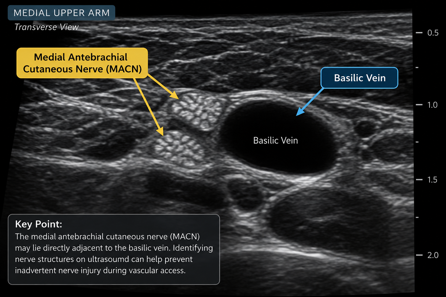
Illustrative ultrasound image depicting the anatomical relationship between the basilic vein and medial antebrachial cutaneous nerve. For educational purposes.
Recognizing Nerves on Ultrasound
Peripheral nerves generally appear as hyperechoic, noncompressible structures. In transverse view, larger nerve branches may demonstrate a characteristic honeycomb appearance, while smaller cutaneous nerves may appear as bright clusters without clearly distinguishable internal features.
These smaller structures can be difficult to differentiate from surrounding connective tissue.
The article encourages clinicians to recognize and avoid unidentified hyperechoic tissue clusters along the proposed needle path rather than focusing exclusively on the target vessel.
Clinical Implications
Historically, clinicians have often relied on patient-reported symptoms, such as sudden shooting or electrical pain, to recognize possible nerve contact.
However, this approach has limitations, particularly among sedated, anesthetized, pediatric, or communication-impaired patients.
The author advocates a more proactive approach: incorporating nerve identification into the routine ultrasound assessment before cannulation.
CCE Clinical Perspective
Successful vascular access requires more than locating a suitable vein. It requires understanding the anatomical relationships between vessels, nerves, and surrounding tissues.
The article reinforces the importance of comprehensive sonoanatomy education and thoughtful vessel selection. Developing the ability to recognize potential hazards before needle insertion is an essential component of safer ultrasound-guided vascular access.
Clinicians can become so focused on identifying a suitable, compressible vein that they overlook surrounding anatomical structures. Recognizing these structures, particularly peripheral nerves, is essential for minimizing the risk of injury, improving the patient experience, and promoting safe vascular access practices.
Understanding the anatomical relationships between vessels and surrounding tissues also allows clinicians to make more informed decisions about insertion sites and needle trajectories, potentially reducing unnecessary attempts and complications.
Plan before you poke!
Reference
Teipen W. Don't Get on My Nerves. Journal of the Association for Vascular Access. 2025;30(1):36–37. https://doi.org/10.2309/JVAD-30-1-Deeper-Dive
